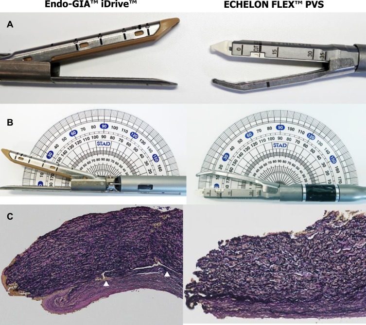
Figure 1.
(A) Comparison of powered endoscopic staplers (B) comparison of jaw angles and (C) histological findings after pulmonary arterial stapling. Histological sections of pulmonary artery tissue adjacent to the area of stapling were stained with Elastica van Gieson stain (X200). In (C) the pulmonary artery section is from stapling with the Endo-GIA™ iDrive™ plus gray cartridge. White arrows indicate the ruptured medial layer.
Abbreviation: PVS, powered vascular stapler.

