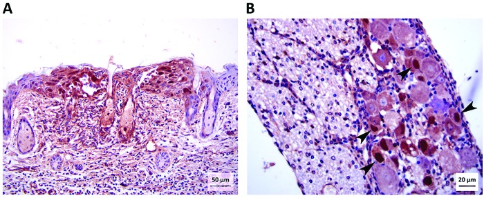
Figure 7.
Photomicrographs of (A) skin (magnification, 200×) and (B) lumbar DRG (magnification, 400×) from an untreated mouse at 3 dpi. Immunohistochemical stain for BV antigen shows a focus of keratinocytes with abundant intranuclear positive signal within the epidermis (skin from inoculation site). A few cells within sebaceous glands near the center of the photo have intranuclear viral antigen. Immunohistochemical staining for BV antigen also shows strong positive signal within the nuclei (arrowheads) of several neurons within the DRG.

