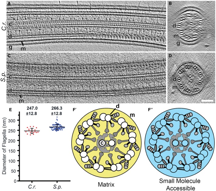
Figure 2. Determination of flagellar volumes.

-
A–DLongitudinal (A and C, proximal view on left) and cross‐sectional (B and D, proximal‐to‐distal view) tomographic slices of representative intact flagella from wild‐type Chlamydomonas reinhardtii (C.r.) flagella (A and B) and sea urchin Strongylocentrotus purpuratus (S.p.) sperm (C and D). Note that in Chlamydomonas, the flagellar membrane (m) is surrounded by a glycocalyx (g). Other labels: central pair complex (c) and doublet microtubule (d). Scale bar, 100 nm.
-
EDiameter of intact flagella from Chlamydomonas and sea urchin sperm measured on cross‐sectional views of cryo‐tomographic reconstructions. Numbers indicate average diameters ± SD (n = 84 for sea urchin (S.p.) and n = 18 for Chlamydomonas (C.r.)).
-
F′, F″Schematic diagrams of Chlamydomonas flagella in cross‐sectional view. The yellow area (F′) indicates the matrix volume V ma that is accessible to proteins, and the cyan area (F″) represents the volume that is accessible to small molecules V asm (i.e., the combination of the matrix volume and the microtubule lumen).
