Figure 2.
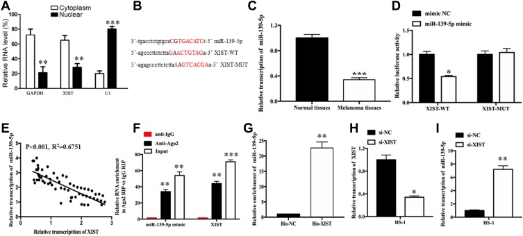
XIST inhibits miR-139-5p expression by acting as a molecular sponge in melanoma cells.
Notes: (A) Relative RNA level of XIST in cytoplasm and nuclear. (B) Binding sequences between XIST and miR-139-5p. (C) Relative levels of miR-139-5p in melanoma tissues and normal tissues. (D) Negative correlation between XIST expression and miR-139-5p expression in melanoma tissues. (E) Luciferase activity in HS-1 cells co-transfected with XIST-wt or XIST-mut reporter and miR-139-5p mimic or miR-NC. (F) RIP assays were performed using ago2 antibody or control IgG antibody in HS-1 cells. (G) RNA pull-down assay was conducted to assess binding between XIST and miR-139-5p in HS-1 cells. (H, I) qRT-PCR analysis of XIST and miR-139-5p expressions in HS-1 cells after transfection with si-NC or si-XIST. Bio-NC, a biotinylated lncRNA that is not complementary to miR-139-5p was employed as a negative control. *P<0.05, **P<0.01, ***P<0.001.
Abbreviations: mut, mutant; qRT-PCR, quantitative reverse transcription-polymerase chain reaction; RIP, RNA immunoprecipitation; si-NC, scrambled RNA for negative control; si-XIST, small nucleolar RNA XIST; WT, wild type; MUT, mutant type.

