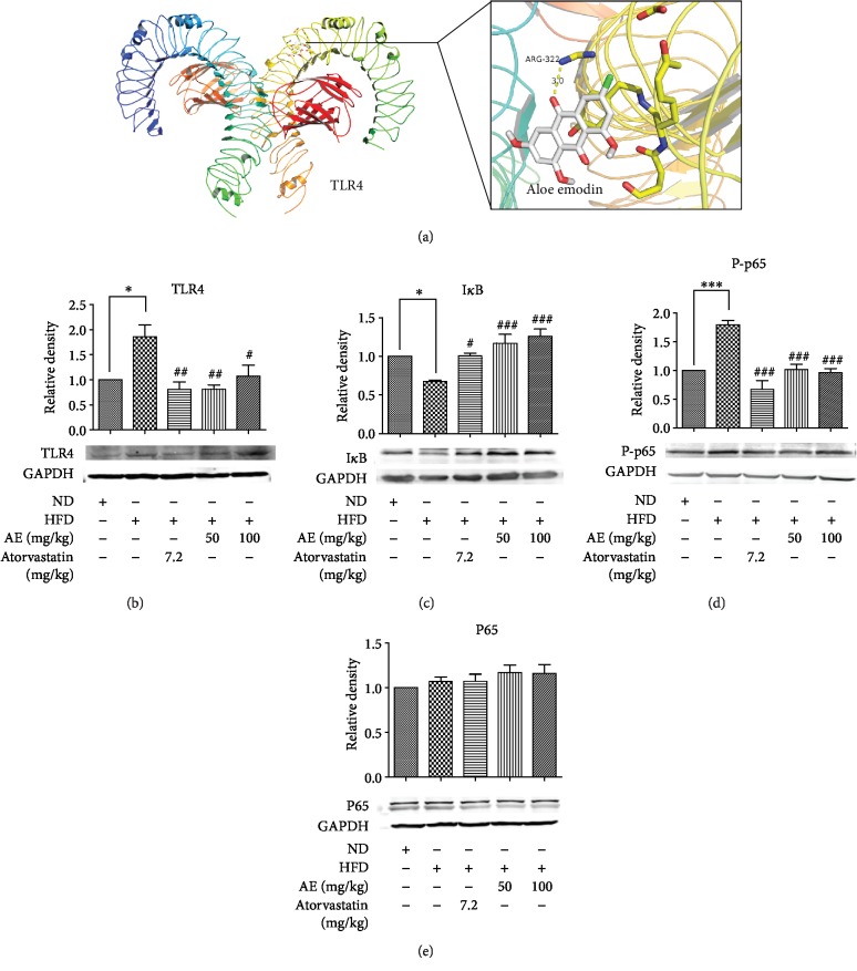
Figure 4.
AE has an important repressor effect on the HFD-stimulated TLR4 pathway signaling. 3D schematic diagram of interaction between AE and TLR4 active site residues: the yellow dotted line is hydrogen bonding (a). Heart tissue homogenate was extracted for western blot assay. GAPDH was used as a loading control. The column figures have shown the normalized optical density of each protein as follows: (b) Toll-like receptor 4 (TLR4), (c) inhibitor of NF-κB (IκB), (d) nuclear factor-kappa B P65 (NF-κB P65), and (e) p-nuclear factor- kappa B P65 (p-NF-κB P-P65) (n = 5; ∗P < 0.0 and ∗∗∗P < 0.001 compared to the ND group; #P < 0.05, ##P < 0.01, and ###P < 0.001 compared to the HFD group).

