
Figure 4.
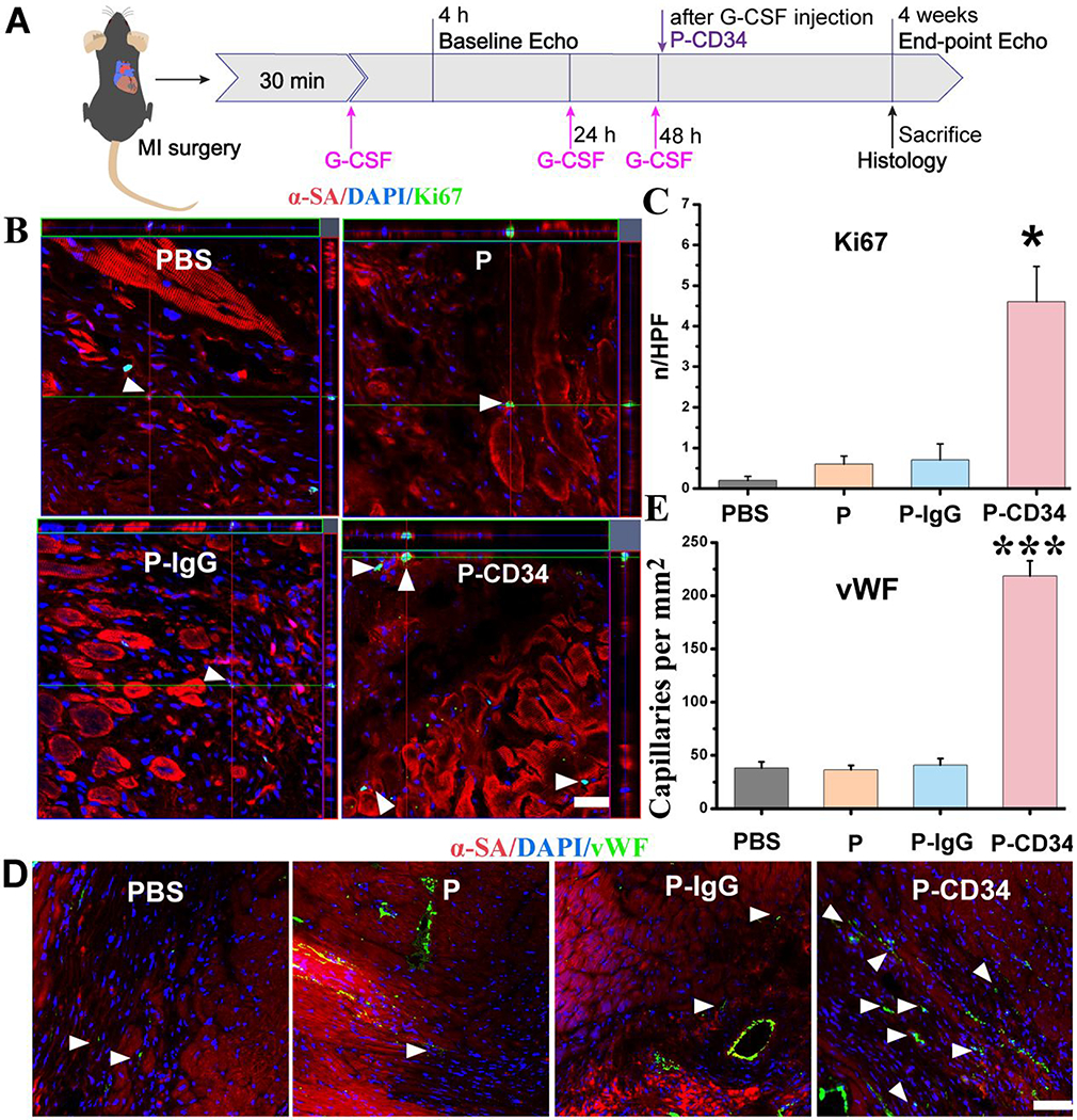
P-CD34 promotion of cardiac repair in a mouse model of heart attack. (A) A schematic showing the animal study design. (B) Z-stack analysis showing Ki67+ nuclei are embedded within cardiomyocytes treated by PBS, platelets, IgG-modified platelets, or CD34-attached platelets 4 weeks later. White arrowheads indicate the positively stained cells. (C) Quantitative analysis of Ki67-positive nuclei. (D) Representative micrographs showing vWF-labeled blood vessels (green) in sample groups (control PBS and hearts treated with platelets, IgG-modified platelets, or CD34-attached platelets) 4 weeks later. White arrowheads indicate the positively stained cells. White arrowheads indicate vWF-positive capillary density in the infarcted area. (E) Quantitative analysis of vWF-positive vasculatures. Scale bar: 20 μm, P-CD34 groups vs other groups. Single asterisks indicate P < 0.05, and triple asterisks indicate P < 0.005.