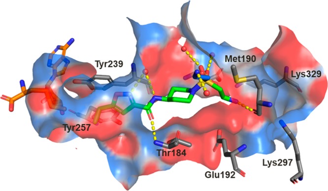
Figure 2.

Crystal structure of compound 3 (green carbons) bound to SMYD3. A second conformation of the ethylamine of compound 3 observed in the crystal structure is colored with blue carbons, the cofactor SAM is colored with orange carbons, and crystallographic waters are represented as red spheres. Key hydrogen bond interactions are depicted with yellow dashes, and the binding site surface is colored by electrostatic potential. An alternate conformation observed for the side chain of Glu192 has been omitted for clarity.
