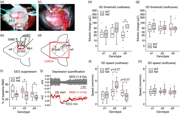
Figure 5.
Na+/K+-ATPase α isoform deficiency and anesthesia differentially affect SD threshold and speed. (a–b) Urethane/α-chloralose protocol with open cranial window preparation and experimental setting. LDF: laser Doppler flowmetry probe; LASCA: laser speckle contrast analysis imaging area; ref: reference electrode; ECoG: electrocorticography electrode; perf: ACSF perfusion; stim: site of electrical stimulation; ECoG: epidurally placed electrode (c–d) Isoflurane protocol with LASCA imaging through the intact mouse skull. (e) In agreement with acute brain slice experiments under normal [K+]o, α2+/KOE4 mice do not display a lower SD threshold. Mice deficient for the α3 isoform required a higher threshold charge to trigger SD. Y-axis shows electric charge in micro Coulomb of logarithmically transformed data. (f) In α2+/KOE4 mice, SD speed showed a tendency towards higher values, whereas the speed tended to be lower in α3+/KOI4 animals. Both observations did not reach statistical significance. (g–h) α isoform deficiency had no effect on SD threshold and speed under isoflurane anesthesia. (i) Depression of spontaneous activity following SD was more pronounced in heterozygous knock-out mice of all lines (α1, α2, α3) compared to their wild-type littermates. Differences reached statistical significance (P < 0.05) in α2-het and α3-het animals (α2: P = 0.04, α3: P = 0.03). (j) Example of spontaneous activity depression with subsequent recovery. EEG suppression was calculated as RMS amplitude reduction of the bandpass-filtered (1–4 Hz). RMS: root mean square. Vo: extracellularly recorded voltage.

