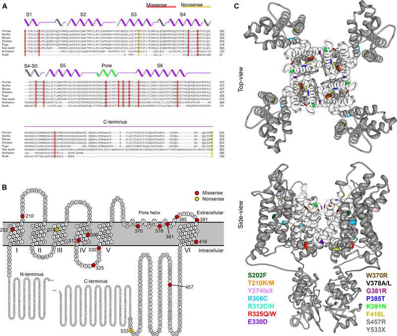
Figure 1. KCNB1 variants identified in individuals with DEE.
A. Evolutionary conservation of KV2.1 shown by multiple sequence alignment of KV2.1 species orthologues (Clustal Omega 39); secondary structural elements are illustrated above the sequences. KV2.1 variants are shaded in red (missense) and yellow (nonsense). B. Schematic view of the entire KV2.1 subunit and the variants in the membrane: modified from Protter plot of Q14721 (KCNB1_Human)40. C. Locations of variants (color-coded) mapped onto crystal structure of KV2.1/Kv1.2 chimera (PDB2R9R)21: a top-view and a side-view across lipid bilayer. Pore domain is highlighted in white; two variants (S457R and Y533X) are not depicted, because their location in the distal C-terminus is not available in crystal structure.

