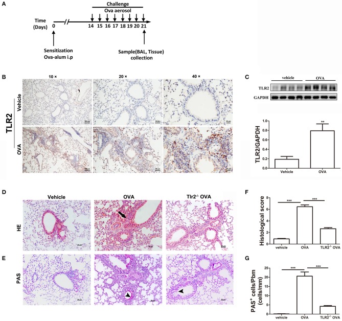
Figure 1.
TLR2 is required for OVA-induced murine allergic airway inflammation. (A) Protocol of establishing allergic airway inflammation, and comparison of resolution of WT and TLR2−/− mice. (B) The expression of TLR2 in lung tissue from vehicle and OVA-challenged mice was analyzed by immunohistochemistry. (C) The protein expression of TLR2 in OVA-challenged WT mice analyzed by western blot and quantification of the protein expression of TLR2. (D) Histological evaluation of the airway inflammation by staining lung sections with H&E, arrows indicates infiltrated leukocytes. (E) Histological examination of mucus production in the lung sections stained with PAS, arrow heads indicates goblet cells. (F) Quantitative analysis of airway inflammation. (G) Quantitative of mucus production. Scale bar: 50 μm. **p < 0.01, ***p < 0.001.

