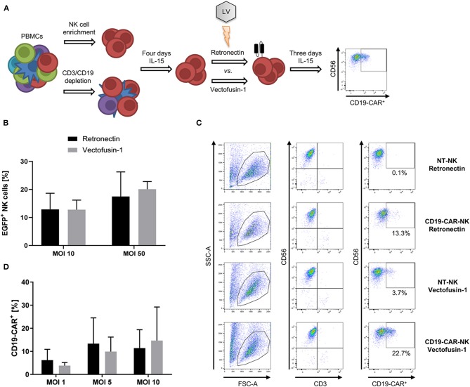
Figure 1.
Comparative lentiviral transduction of NK cells using Retronectin and Vectofusin-1. (A) Scheme of the transduction procedure. NK cells were isolated from PBMCs either by NK cell enrichment or CD3/CD19 depletion and cultivated for 4 days with IL-15 prior to transduction comparing the enhancers Retronectin and Vectofusin-1. After transduction, NK cells were cultivated for 3 days with IL-15 until transduction efficiency was analyzed by flow cytometry. (B) NK cells from three donors (n = 3) were transduced with VSV-G pseudotyped lentiviral EGFP particles at two different multiplicities of infection (MOI) and with two different transduction enhancers. (C) Gating strategy to estimate the transduction efficiency of NK cells transduced with VSV-G pseudotyped lentiviral CD19-CAR particles (e.g., for more detailed gating strategy see Supplementary Material). NK cells were identified as CD56+CD3− leukocytes (first and second column). From those CD19-CAR+ NK cells were estimated (third column). In the first and second row representative data of NK cells are depicted that were transduced with Retronectin at MOI 5 vs. non-transduced (NT) NK cells from NK cell preparations of the same donor. In the third and fourth row data from NK cells transduced with Vectofusin-1 at MOI 5 vs. NT-NK cells are shown. Percentage of false positive CD19-CAR events in NT-NK cells was subtracted from the percentages measured in the belonging transduced NK cells. Shown are the dot plots of one donor. (D) NK cells from four donors (n = 4) were transduced with VSV-G pseudotyped lentiviral CD19-CAR particles at shown MOIs and with two different transduction enhancers. Shown are mean values + SD. Statistical analysis was performed using two-tailed student's paired t-test. No significant differences were observed between analyzed groups in (B,D) as p-values were >0.05.

