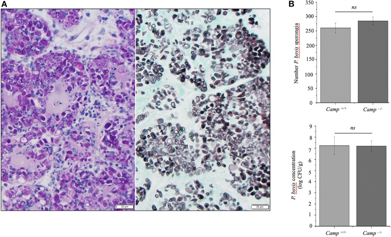
Figure 2.
Prototheca bovis was present at similar levels in the mammary glands of Camp+/+ and Camp−/− mice. (A) Mammary gland from a Camp+/+ mouse infected with P. bovis (4 d post-infection). Abundance of Prototheca organisms within tissue sections was revealed with special stains. Prototheca are stained magenta by periodic acid-Schiff (PAS) stain (left image) and black by Grocott methenamine silver (GMS) stain (right image). Scale bar = 20 μm. (B) P. bovis was present in mammary glands at similar levels in Camp+/+ and Camp−/− mice, as determined by counting of sporangia per field (minimum of 10 randomly chosen microscopic fields, x40 objective magnification) and culturing 10-fold diluted homogenates on Sabouraud dextrose agar. Data are means ± SEM (n = 4/group). Ns, not significant.

