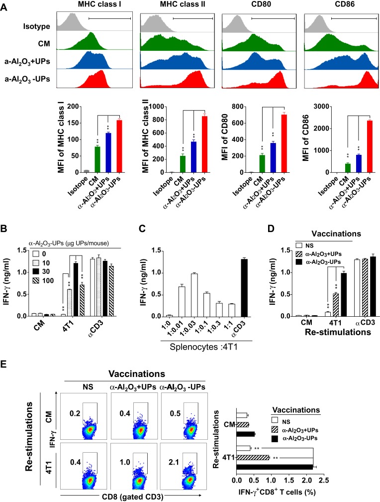
Figure 4.
Effect of α-Al2O3-UPs on the expression of MHC class I, MHC class II, CD80, and CD86 molecule on DCs and tumor-specific immune response induced by α-Al2O3-UPs. (A) Expression analysis of MHC class I, MHC class II, CD80 and CD86 on BMDCs by flow cytometry. Cultured BMDCs were treated for 48 h with CM (green area), α-Al2O3 +UPs (blue area) or α-Al2O3-UPs (red area). (B) BALB/c mice (n=3 per group) were vaccinated with 0, 10, 30 or 100 μg α-Al2O3-UPs. The splenocytes from the vaccinated mice were then re-stimulated with inactivated 4T1 tumor cells or without stimulation (CM). αCD3-Ab stimulation was used as a positive control. IFN-γ secretion was detected by ELISA. (C) Splenocytes from the vaccinated mice were re-stimulated with different numbers of inactivated 4T1 tumor cells and the levels of IFN-γ were measured by ELISA. (D) BALB/c mice (n=3 per group) were vaccinated with NS, α- Al2O3+UPs (30μg) or α- Al2O3-UPs (30μg) via triple subcutaneous injections. The IFN-γ production by the responder cells was detected after 48 hrs by ELISA. (E) Flow cytometric analysis of intracellular IFN-γ produced by CD8+ T cells. Data (means ± SD) are representative of three independent experiments results. **, P < 0.01; ns, not significant, by One-way ANOVA with the Tukey-Kramer multiple test, two-tailed unpaired t-test or Mann–Whitney U-test.

