
Fig. 5.13.
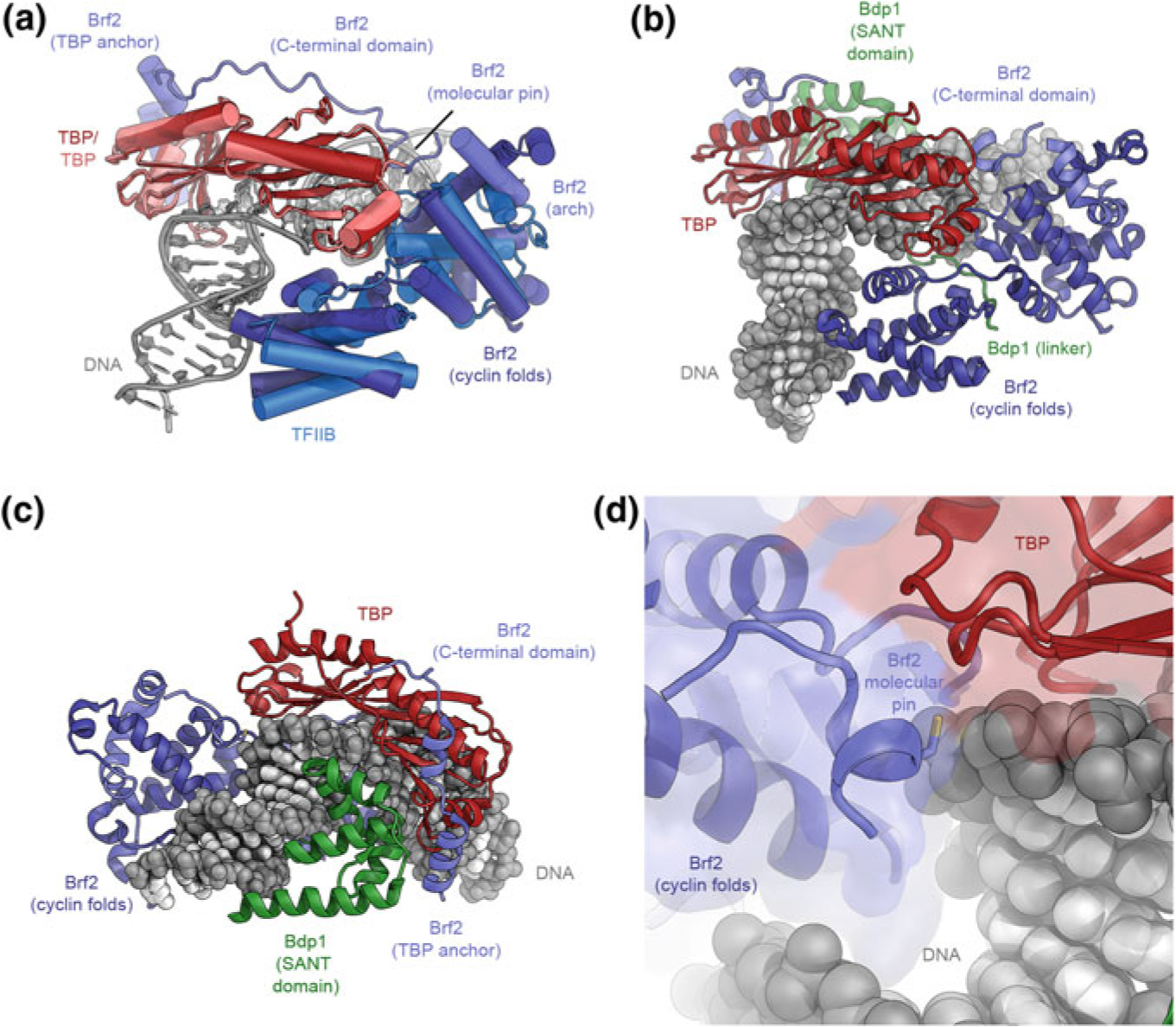
The structure of TFIIIB. a Superposition of the structures of the DNA-bound TBP-Brf2 complex (Gouge et al. 2015) and the TBP-TFIIB-DNA complex (Tsai and Sigler 2000). TBP and the Brf2 cyclin folds in TFIIIB occupy very similar positions as their Pol II-system counterparts (TBP and TFIIB, respectively). The C-terminal domain of Brf2 comprises arch, molecular pin, and TBP anchor regions (as indicated). b, c Structure of DNA-bound TFIIIB (Gouge et al. 2017). The Bdp1 SANT domain binds to DNA at the side opposite to the Brf2 cyclin folds. d Structure of the redox-sensitive molecular pin in the context of bound DNA and TBP (Gouge et al. 2017)