
Fig. 5.15.
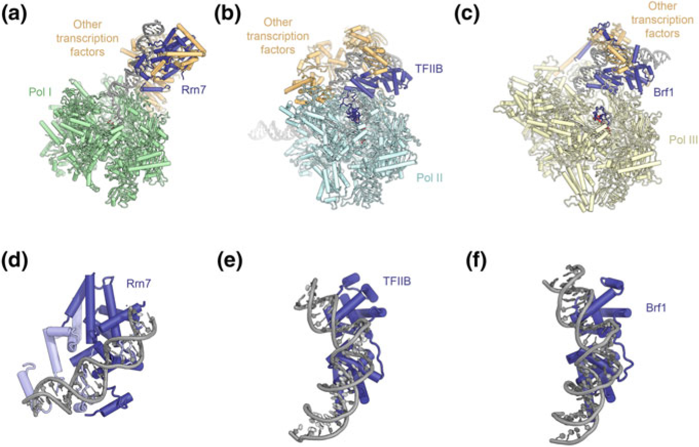
Comparison of DNA-bound structures of TFIIB-like initiation factors. a–c Structure of the Pol I (a), Pol II (b), and Pol III (c) ITCs (Han et al. 2017; He et al. 2016; Vorländer et al. 2018). TFIIB-related factors are colored dark blue, other transcription factors orange. d–f Conformation of upstream promoter DNA bound to the cyclin folds of Rrn7 (d), TFIIB (e), and Brf1 (f)