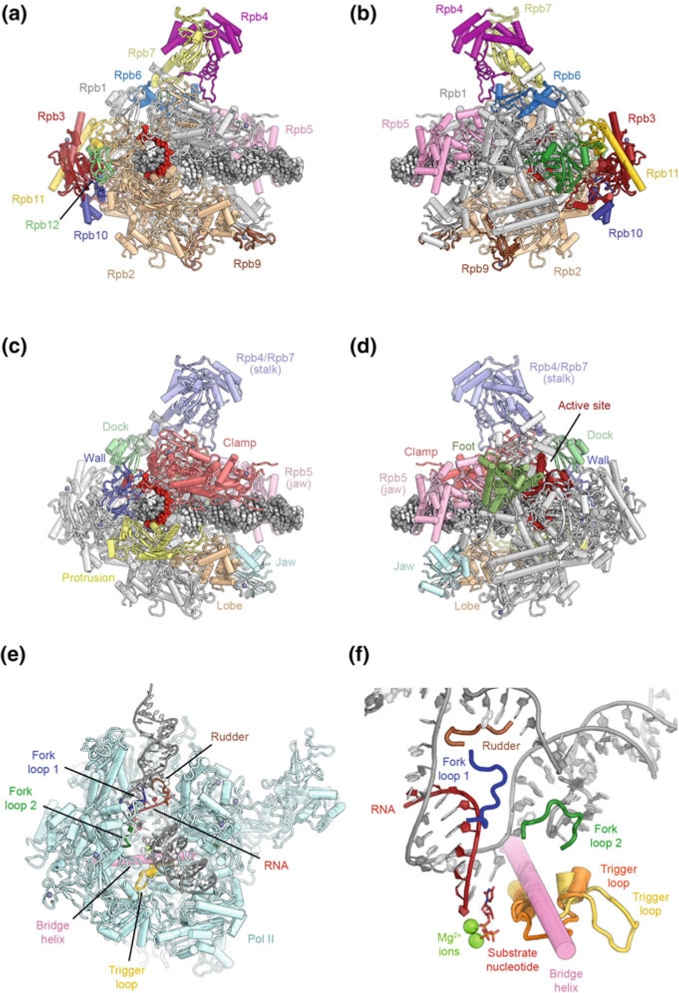
Fig. 5.2.

Architecture of eukaryotic RNA polymerase II. a, b Depiction of transcribing Pol II extracted from the structure of an elongation complex (Ehara et al. 2017), shown from the front and back and colored by protein subunits. c, d Structural domains of the Pol II subunits Rpb1 and Rpb2 are highlighted in color according to (Cramer et al. 2001). Additional structural landmarks mentioned in the text are highlighted as well (protein names are indicated). e View of the transcription bubble at the Pol II active site (Barnes et al. 2015). Key structural and functional elements near the active site that are mentioned in the text are indicated. f Detailed view of the Pol II active site (Barnes et al. 2015; Wang et al. 2006). Two conformations of the trigger loop are shown in orange and yellow
