
Fig. 5.3.
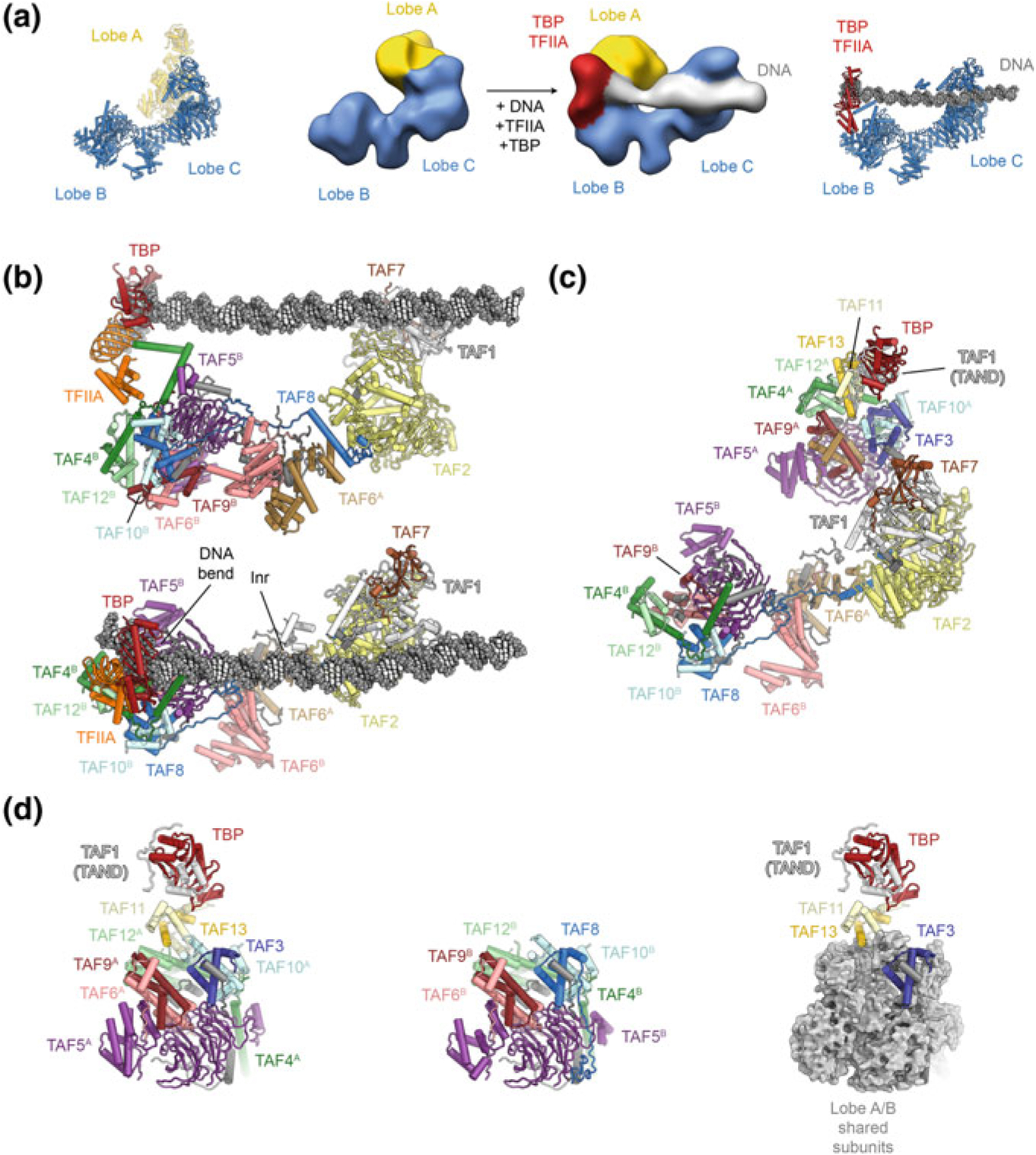
The structure of human TFIID. a Organization of human TFIID into three lobes. Early cryo-EM maps (Cianfrocco et al. 2013) that revealed the tri-lobal overall structure of TFIID are shown in the center, with recent coordinate models (Patel et al. 2018) shown next to them for comparison. b Structure of TFIID bound to DNA and TFIIA (Patel et al. 2018) in side view (top) and top view (bottom). Protein subunits are shown in colour and labelled; the TBP-induced DNA bend and the location of the transcription start site (Inr) are labeled. c Structure of TFIID in the canonical state (Patel et al. 2018) shown in the same orientation as bottom panel in (b). d Composition of lobes A and B. Left: lobe A. Center: lobe B. Right: Subunits shared between lobes A and B are shown as grey surface to highlight the subunits in lobe A that distinguish between lobes A and B