
Fig. 5.6.
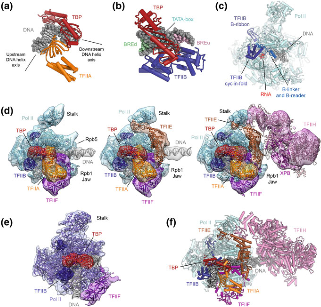
The structure of the Pol II-PIC and its components. a Structure of promoter DNA bound to TBP and TFIIA (Bleichenbacher et al. 2003). TBP introduces a pronounced bend into the TATA box (indicated by dotted black lines representing the upstream and downstream DNA helix axes). b Structure of promoter DNA bound to TBP and TFIIB (Tsai and Sigler 2000). The TATA box and regions corresponding to the BREd and BREu regions recognized by TFIIB are indicated by light blue, green, and pink shading of the DNA bases. c Structure of initially transcribing Pol II bound to TFIIB (Sainsbury et al. 2013). d Stepwise assembly of the human Pol II-PIC: Pol II-DNA-TBP-TFIIA-TFIIB-TFIIF (left), Pol II-DNA-TBP-TFIIA-TFIIB-TFIIF-TFIIE (middle), and Pol II-DNA-TBP-TFIIA-TFIIB-TFIIF-TFIIE-TFIIH (right), the latter showing a bilobal architecture. Depiction based on (He et al. 2013) with fitted coordinates from (Greber et al. 2019; He et al. 2016). e The structure of the yeast Pol II-ITC lacking TFIIH, TFIIA, and TFIIE (Plaschka et al. 2015) is in good agreement with the human Pol II-PIC core (compare e.g. to leftmost complex in panel d). f Complete molecular structure of the human Pol II-PIC with TFIIH, assembled from coordinates for the human core Pol II-PIC and the structure of human TFIIH (Greber et al. 2019; He et al. 2016)