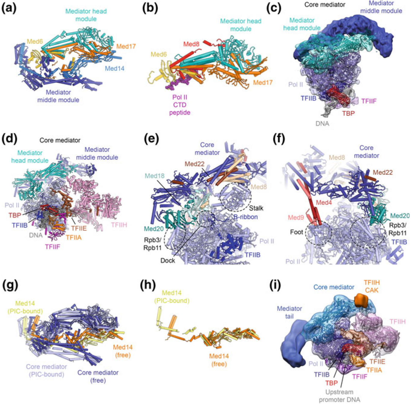
Fig. 5.8.

The Mediator-bound Pol II-PIC. a Crystal structure of the Mediator head and middle modules (core Mediator) (Nozawa et al. 2017). b Crystal structure of the Mediator head module bound to a Pol II CTD peptide (Robinson et al. 2012). c Low-resolution cryo-EM structure of the core-Pol II-ITC bound to core Mediator (Plaschka et al. 2015). d Cryo-EM structure of the yeast Pol II-PIC, including TFIIH, bound to core Mediator (Schilbach et al. 2017). e, f Contacts sites between core Mediator and Pol II, according to the structure shown in (d). g, h Conformational changes in Mediator upon binding to the PIC (g) and associated with conformational changes in Med14 (Nozawa et al. 2017; Schilbach et al. 2017). h Low-resolution cryo-EM map of the yeast Pol-II PIC with Mediator (Robinson et al. 2016), including the Mediator tail module, fitted with the coordinates of the most recent yeast Pol II-core Mediator structure (Schilbach et al. 2017)
