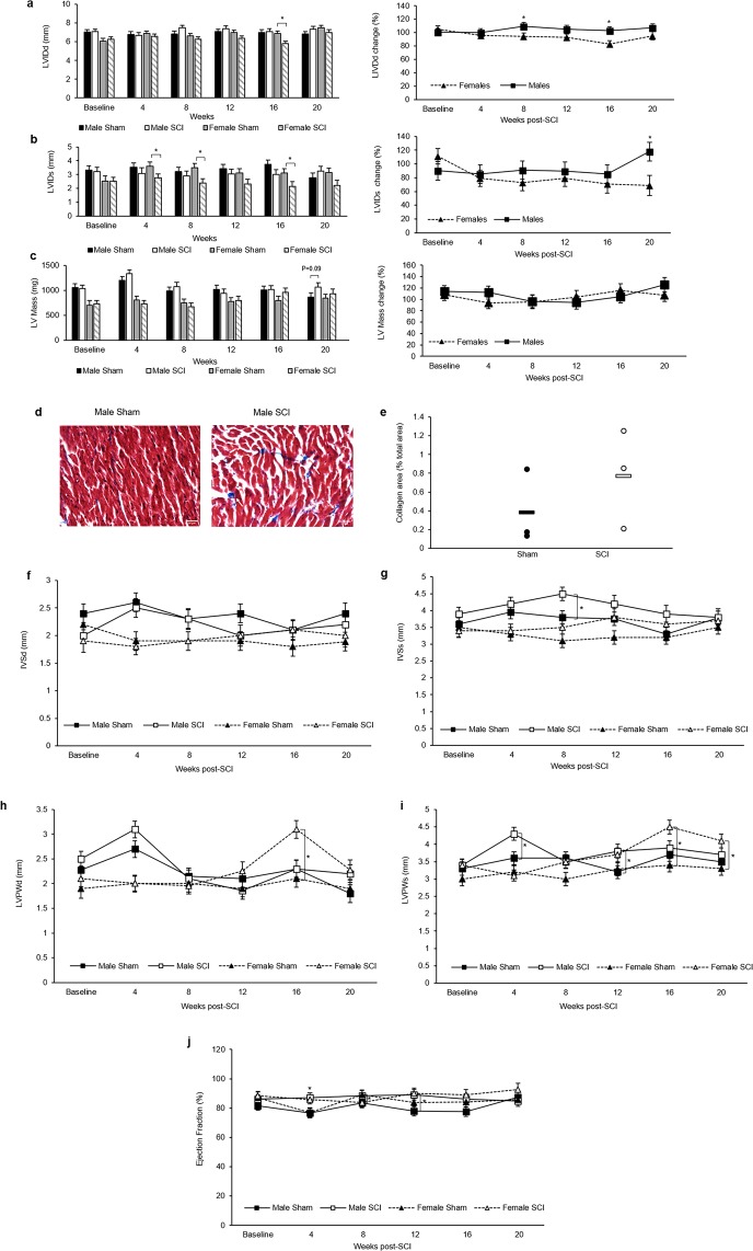
Figure 3.
Echocardiography assessment. (a) Left ventricular internal diameter during diastole (LVIDd) and (b) during systole (LVIDs). (c) LV mass. (d) Representative 40x sections of Masson’s trichrome stained cardiac tissue from male rats. (e) Male collagen area (blue staining) as a % of total tissue area. (f) Intraventricular septum during diastole (IVSd) and (g) during systole (IVSs). (h) Left ventricular posterior wall thickness during diastole (LVPWd) and (i) during systole (LVPWs). (j) Ejection fraction (EF). Statistical analysis was performed using the repeated measures mixed procedure in SAS and data are presented as Least Squares Mean (LSM) ± SEM (*P < 0.05).

