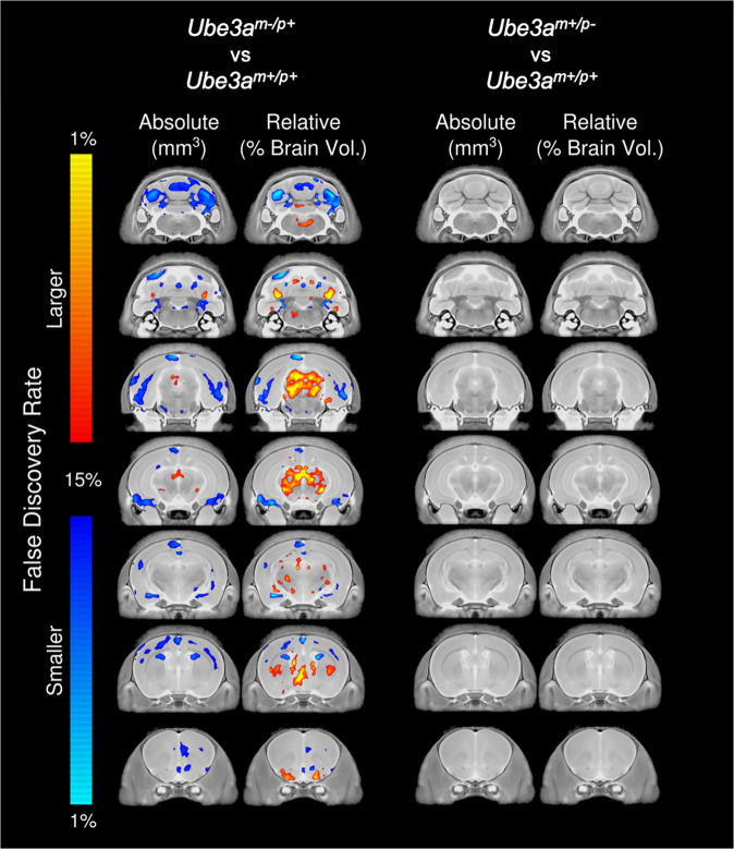
Fig. 4. Neuroanatomical pathology in Ube3am−/p+ rats at PND 21.
Representative coronal slice series highlighting regional brain differences in absolute (mm3) and relative (%) brain volume between Ube3am−/p+ and Ube3am+/p+ (left) and between Ube3am+/p− and Ube3am+/p+ (right). Regions with decreased volume in Ube3am−/p+ include the cerebral cortex, cerebellum, and amygdala. Regions with increased volume in Ube3am−/p+ include the periacqueductal gray, thalamus, and hypothalamus. Ube3am+/p− did not exhibit altered neuroanatomy compared to wildtype. Analyses include both males and females.

