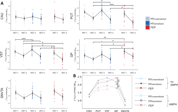
Fig. 2. Dopamine (DA) D2/3 receptor binding ([11C]-(+)-PHNO BPND values) in scans without (PET1 and PET3) and with prior administration of amphetamine (AMPH; PET2 and PET4).
a Panels show binding in five subcortical regions of interest (ROIs; CAU, caudate; PUT, putamen; VST, ventral striatum; GP, globus pallidus; SNVTA, substantia nigra/ventral-tegmental area) in healthy volunteers before (HVUNSENS; sky-blue) and after AMPH sensitization (HVSENS; deep-blue) and in patients with first-episode psychosis (FEP; red). X-axis break between PET2 and PET3 in HV indicates a scan-free interval (2–4 weeks) during prospective AMPH sensitization. All direct AMPH effects (PET1 vs. PET2 HVUNSENS and FEP, PET3 vs. PET4 HVsens) were significant (paired t-tests p = 0.03–2.5 × 10⁻⁵; not marked). Note that AMPH sensitization led to a significant increase in D2/3 receptor binding from PET1 to PET3 in VST. b Alternative representation of data shown in a highlighting systematic differences in D2/3 receptor binding across conditions between HV and patients with FEP. D2/3 binding in FEP is lower than in HV in D2 receptor-rich neo-striatal regions (CAU, PUT, VST), while it is elevated in D3 receptor-rich regions of the paleo-striatum (GP and SNVTA). Error bars represent 95% confidence intervals. *p < 0.05, **p < 0.01, ***p < 0.001, post-hoc two-tailed t-test.

