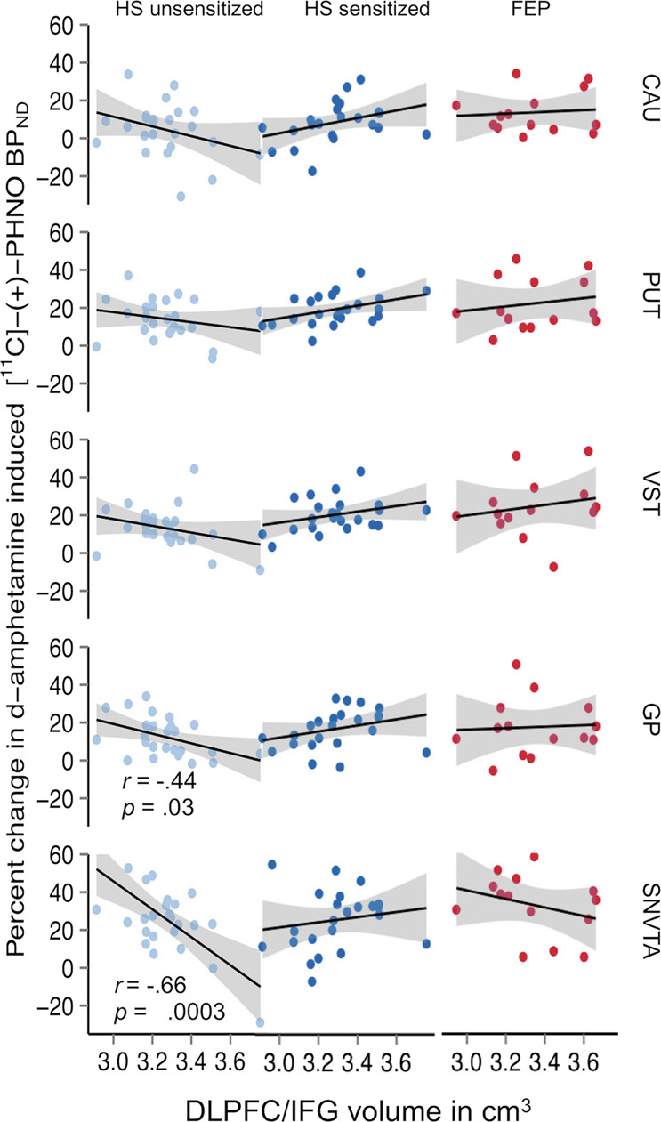
Fig. 5. Relationship between volume of the dorsolateral prefrontal cortex (in cm3) and inferior frontal gyrus (DLPFC/IFG) as measured with magnetic resonance imaging and AMPH-induced reductions in non-displaceable binding potential (BPND) values of the dopamine D2/3 receptor positron emission tomography (PET) radioligand [11C]-(+)-PHNO in healthy subjects before and after sensitization to d-amphetamine compared to patients with schizophrenia.

Significant interactions are found in brain regions (GP, SNVTA) where dopamine D3 (in contrast to D2) receptors are the predominant source of signal detected with [11C]-(+)-PHNO and PET. VST, ventral striatum; GP, globus pallidus; SNVTA, substantia nigra/ventral tegmental area.
