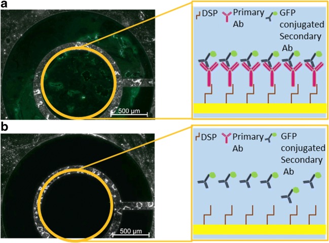
Fig. 2.
Optical characterization of the antibody-modified electrode. Optical images of electrodes incubated with a secondary GFP-conjugated antibody following an incubation with DSP and the primary antibody (a) or only with DSP (b). In the illustration, the electrode is denoted as a yellow square, DSP is denoted as a brown stick, the primary antibody as a pink ‘Y’ shape, and the secondary antibody as a dark blue ‘Y’ shape with a small green circle on its base

