Figure 5.
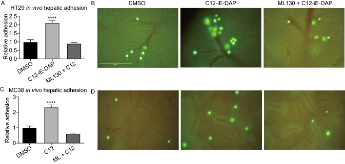
NOD1 stimulation increasesin vivohepatic sinusoid adhesion of circulating tumor cells (CTCs). (A) Relative in vivo adhesion of HT29 sinusoids under DMSO, C12 stimulation (2,000 ng/mL), and ML130 co-incubation (10 μmol/L) (n = 8 mice/condition). NOD1 activation leads to a 2-fold increase in in vivo hepatic adhesion compared to DMSO or ML130 co-incubation (P < 0.0001). (B) Representative microscopy images of HT29 adhesion to hepatic sinusoids under DMSO, C12, and ML130 + C12 are shown. Green intensities represent HT29 cells live-stained with CSFE and the reticular patterns represent hepatic sinusoids. Compared to DMSO (left), C12-induced (middle) NOD1 activation leads to enhanced hepatic sinusoid adhesion. Such enhanced adhesion is absent when co-incubated with ML130 (right). (C) To show a generalized effect of NOD1 activation on hepatic adhesion, the above experiment is repeated using murine colon cancer MC38 (n = 5 mice/condition). The same results are obtained in which C12 stimulated cells (middle) showed 2-fold increase in in vivo adhesion. (D) Representative images of MC38 adhesion to hepatic sinusoids are shown. (E) Scale bar measures 1mm and applies to all microscopy images in Fig. 5. Error bars represent SEM. All comparisons are made with respect to the DMSO control. Only significant comparisons are labelled. “***” denotes P < 0.0001

