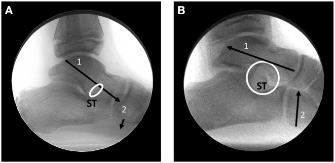
Figure 5.

Lateral weight bearing fluoroscopic radiographs. (A) Relaxed stance position. The talus has partially dislocated on the calcaneus (1). The sinus tarsi (ST) is obliterated and the navicular has dropped (2). (B) The same foot with the articular facets of the talus repositioned on the calcaneus (1). Sinus tarsi (ST) is re-opened and the navicular is elevated (2).
