
Fig. 2.
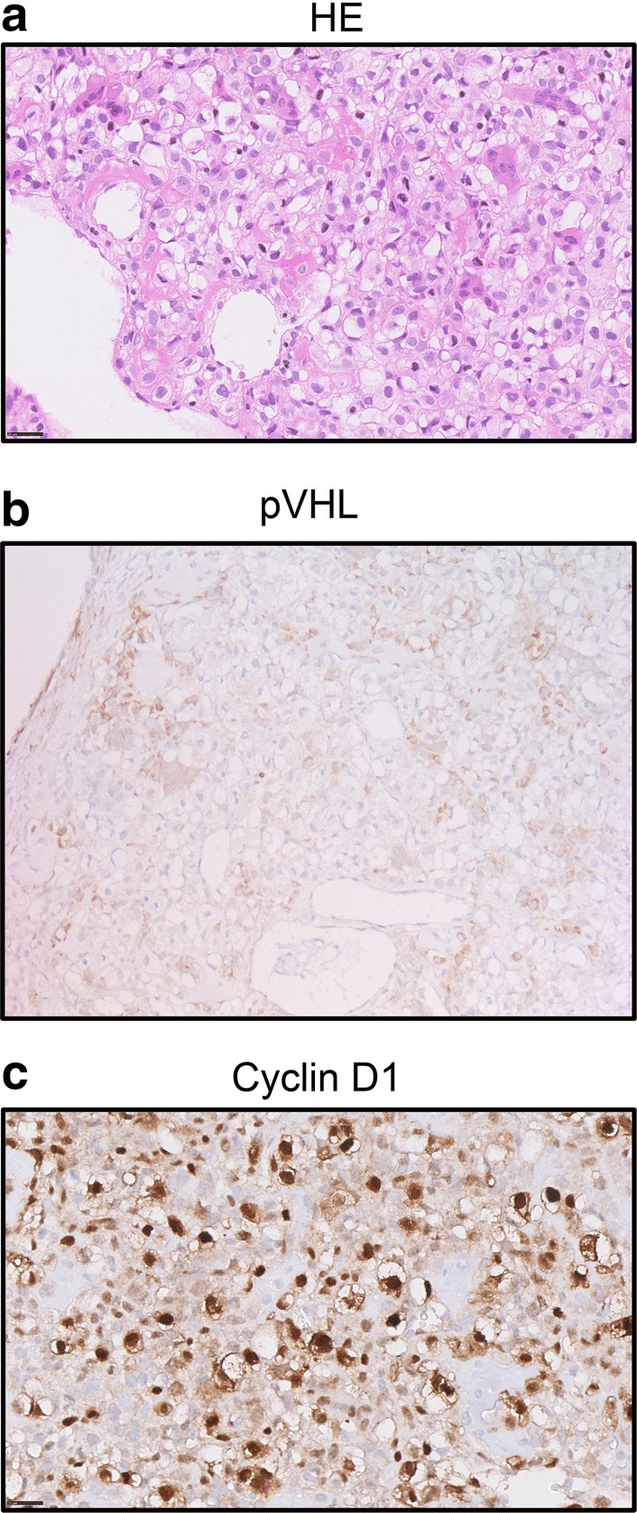
Molecular analysis of the tumor after surgical removal of the clear cell chondrosarcoma. a Hematoxylin and eosin (HE) stain of the tumor tissue (× 40 magnification). The tumor is composed of cells with distinct borders and clear to slightly eosinophilic cytoplasm with central nuclei. Osteoclast-type giant cells, reactive bone and wide vessels are present in the tumor. b Immunohistochemical staining of the tumor tissue using antibodies directed at pVHL (20 ×). Positive staining is observed in non-neoplastic multinucleated giant cells and other histiocytic cells present in the tumor, which are CD68 positive (not shown). c Immunohistochemical staining using antibodies directed at Cyclin D1 shows abundant expression in the tumor cells (×40)