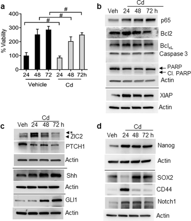
Fig. 1. Effect of acute Cd exposure on BPH1 cell proliferation, Shh signaling activation, and stem cell markers in a time-dependent manner.

a BPH1 cells were exposed to Cd for 24, 48, and 72 h; cell proliferation was determined by MTT assay. b Cells were incubated with or without 10 µM Cd for the indicated time. Cell lysates were subjected to western blotting using antibodies against p65, Bcl2, BclxL, XIAP, cleaved caspase-3, cleaved PARP, and β-actin was used as loading control. c Total cell lysates extracted from cells exposed to Cd (24, 48, and 72 h) were subjected to western blot analysis using specific antibodies against Shh signaling. d Cell lysates from cells exposed to Cd (24, 48, and 72 h) were used to determine the protein level of Nanog, SOX2, CD44, and Notch1 by western blot analysis. β-actin was used as the loading control. #, Not significant; *, nonspecific band.
