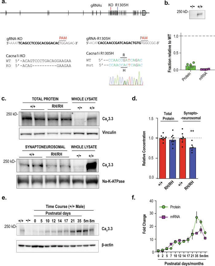
Fig. 1. Generation and biochemical characterization of knock-in and knock-out animals.
a The exon/intron structure of mouse Cacna1i gene, and the positions of targeting sgRNAs for generating the CaV3.3 knock-out (KO) and R1305H knock-in animals. The sequences of the targeting sgRNAs and the genomic sequence verification for knock-out and knock-in founder animals are shown underneath. b Western blot from Cacna1i−/− and Cacna1i+/+ whole-brain lysates showing a near complete lack of CaV3.3 protein in the Cacna1i−/− (−/−) samples. Significant reductions of both mRNA and protein levels in the heterozygous KO mice were also found (Supplementary Fig. 1b–d). Bottom panel: quantification of CaV3.3 protein and mRNA levels normalized to Cacna1i+/+ levels (n = 5). c A typical representative western blot showing total CaV3.3 protein levels (top panel) and CaV3.3 protein levels in crude synaptoneurosomal preparations (bottom panel) from brain tissues derived from littermates of Cacna1i+/+ (+/+) and Cacna1iRH/RH (RH/RH). d Quantification of total and synaptoneurosomal CaV3.3 levels in the total brain lysate (n = 8) and crude synaptoneurosomal preparation (n = 6). e A typical western blot showing the developmental progression of CaV3.3 expression in brain tissue across different postnatal days, and at 5 (5m) and 8 months (8m). f Quantification of protein (n = 2 for each time point) and mRNA (n = 2 for each time point except for 5m and 8m that contains n = 3 each) levels during developmental progression. **p < 0.01; error bars represent S.E.M.

