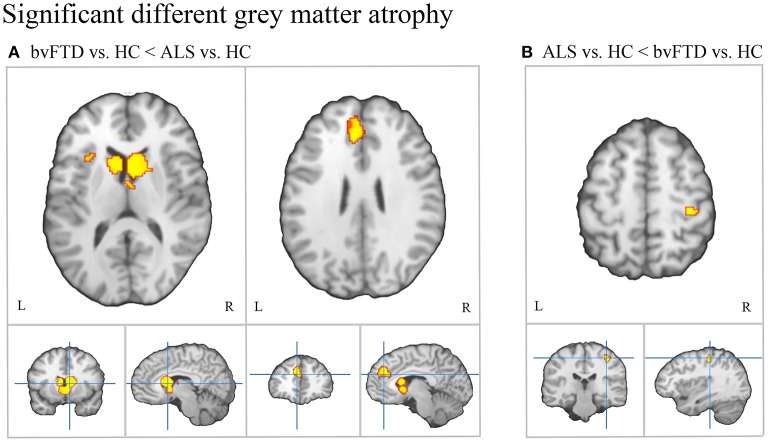
Figure 4.
Patterns of distinct gray matter atrophy between bvFTD and ALS. (A) Significantly greater gray matter atrophy in the medial frontal/anterior cingulate cortex, bilateral caudate, and left anterior insula in bvFTD relative to ALS. (B) Significantly greater gray matter atrophy in the right motor cortex in ALS relative to bvFTD.

