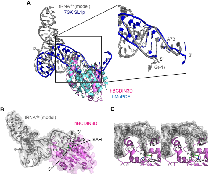
Figure 4.
A model of tRNA docking onto hBCDIN3D. (A) Docking of tRNAHis onto hBCDIN3D. Superimposition of hBCDIN3D (purple) on the structure of the MTD of hMePCE (cyan) complexed with 7SK SL1p (blue). tRNAHis (gray) was modeled such that the acceptor helix of tRNAHis superimposed well onto the 5′-end and 3′-single-stranded region of SL1p. See ‘Materials and Methods’ section for details. (B) α6′ would interact with the major groove of the acceptor helix of tRNAHis, and α0 would wedge the G−1:A73 mispair of tRNAHis. (C) A detailed stereo view of the interaction between the acceptor helix of tRNAHis and BCDIN3D in (B). tRNA is shown as a gray stick model.

