Figure 6.
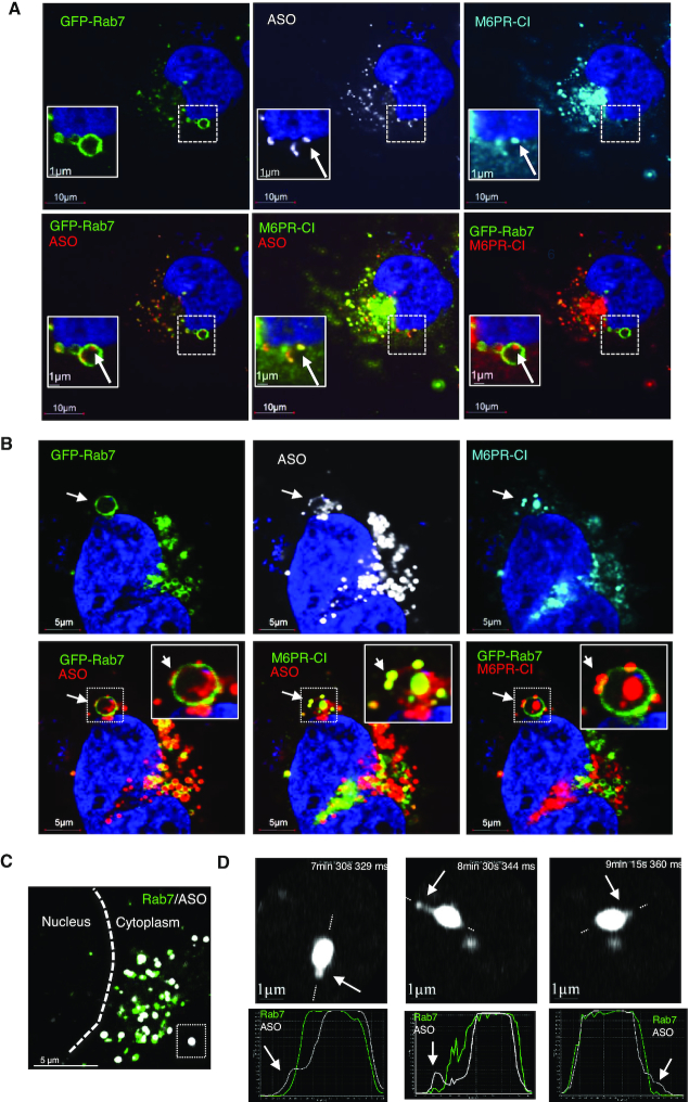
M6PR-CI co-localizes with PS-ASOs inside LEs and on the membranes of LEs. (A) Immunofluorescent staining of M6PR-CI in GFP-Rab7-expressing SVGA cells incubated with 2 μM PS-ASO 446654 for 12 h. Scale bars, 10 μm. The regions boxed with dashed lines are shown at higher magnification in insets. M6PR-CI co-localization with a distinct PS-ASO-containing focus inside the LE is marked with an arrow. (B) Immunofluorescent staining of M6PR-CI in GFP-Rab7-expressing SVGA cells incubated with 2 μM PS-ASO 446654 for 12 h. Co-localization of M6PR and PS-ASOs as a distinct structure on the LE membrane is marked by arrows. Scale bars, 5 μm. (C) Live cell imaging of a SGVA cell incubated with PS-ASO 446654 for 6 h. The nuclear border is marked by a dashed line. The boxed PS-ASO-containing LE in the lower right is magnified in panel D. (D) Upper images: Snapshots taken during live cell imaging. Potential PS-ASO release events are marked by arrows. Lower plots: Signal intensity profiles for the LE across the lines as indicated in the upper panels. Arrows indicate ASOs outside the LE body.

