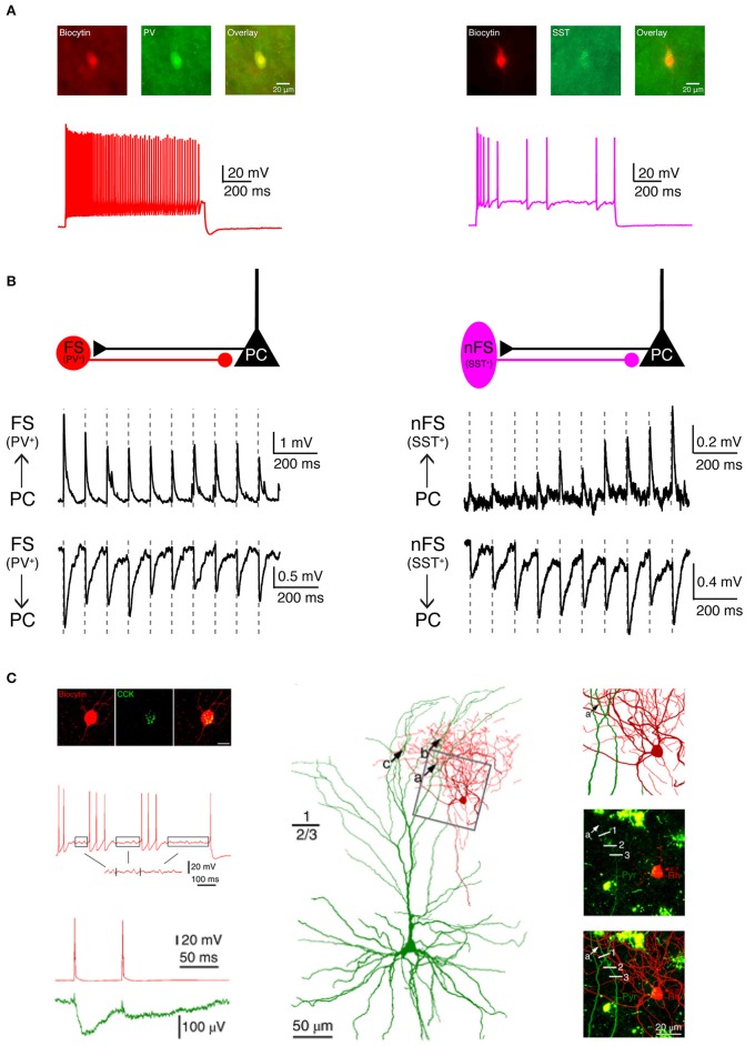
Figure 3.
Electrophysiological, morphological and molecular characterization of synaptic connections by combining paired recordings with immuno-fluorescent stainings for specific marker proteins. (A) Two main types of GABAergic interneurons in the neocortex are PV+ fast spiking interneurons (left, red) which express the Ca2+-binding protein parvalbumin (PV) and SST+ non-fast spiking interneurons (right, violet) which express the neuropeptide somatostatin (SST). (B) Two interneuron types form synaptic connections with different characteristics. Left, PV+ fast spiking interneurons receive initially strong but quickly depressing EPSPs from neighboring excitatory neurons. At the same time, they produce depressing IPSPs in synaptically connected neighboring excitatory neurons. Right, SST+ non-fast spiking interneurons, in contrast, receive initially weak and gradually facilitating EPSPs from neighboring excitatory neurons and in turn elicit facilitating IPSPs in their target excitatory neurons. (C) Boldog et al. identified a specialized human cortical GABAergic cell type, the so-called L1 rosehip cell (RC). L1 RCs express cholecystokinin (CCK), but not PV, SST, or other molecular markers. L1 RCs exhibit an intermittent non-fast spiking firing pattern with subthreshold membrane potential oscillations (boxed segments). By combining paired recordings with Ca2+ imaging the authors were able to demonstrate that L1 RCs establish inhibitory synapses onto apical dendritic tufts of L2/3 pyramidal cells to regulate the AP backpropagation in a segment-specific manner. Electrical signals and morphologies of L1 RCs are in red and those of L2/3 pyramidal cells in green. (A,B) have been adapted from Feldmeyer et al. (2018) with permission and (C) from Boldog et al. (2018) with permission.

