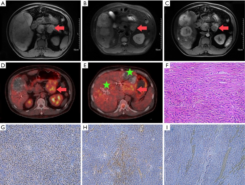
Figure 1.
Imaging, histopathological, and immunohistochemical findings. (A,B,C) Magnetic resonance imaging: axial T1-weighted imaging shows a well-circumscribed mass in the pancreatic head (red arrow), with decreased signal intensity relative to that of the normal pancreas (A); axial T2-weighted imaging shows the mass (red arrow) with heterogeneous, slightly high signal intensity relative to that of the normal pancreas (B); axial contrast-enhanced imaging shows the mass (red arrow) with heterogeneous increased arterial phase enhancement (C); positron emission tomography/computed tomography (D,E): the pancreatic mass can be observed with significantly different degrees of FDG uptake (D; red arrow; SUVmax: 4.74). Multiple, slightly hypodense lesions exhibiting incomplete lipiodol uptake (E; green stars) and remarkable levels of FDG uptake (E; red arrow; SUVmax: 4.79–5.04) are present in the liver; (F,G,H,I) histopathological and immunohistochemical findings: the mass is a spindle cell tumor rich in heterotypic cells (400×) (F); immunohistochemistry shows positive staining for STAT6 (200×) (G), CD34 (200×) (H), and Bcl-2 (200×) (I). SUVmax, maximum standardized uptake. FDG, fluorodeoxyglucose.

