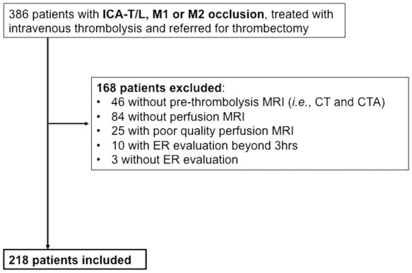
Figure 1.

Study flow chart.
CT: computerized tomography; ER: early recanalization; ICA-T/L: intracranial internal carotid artery occlusion; M1: first segment of the middle cerebral artery; M2: second segment of the middle cerebral artery; MRI: magnetic resonance imaging.
