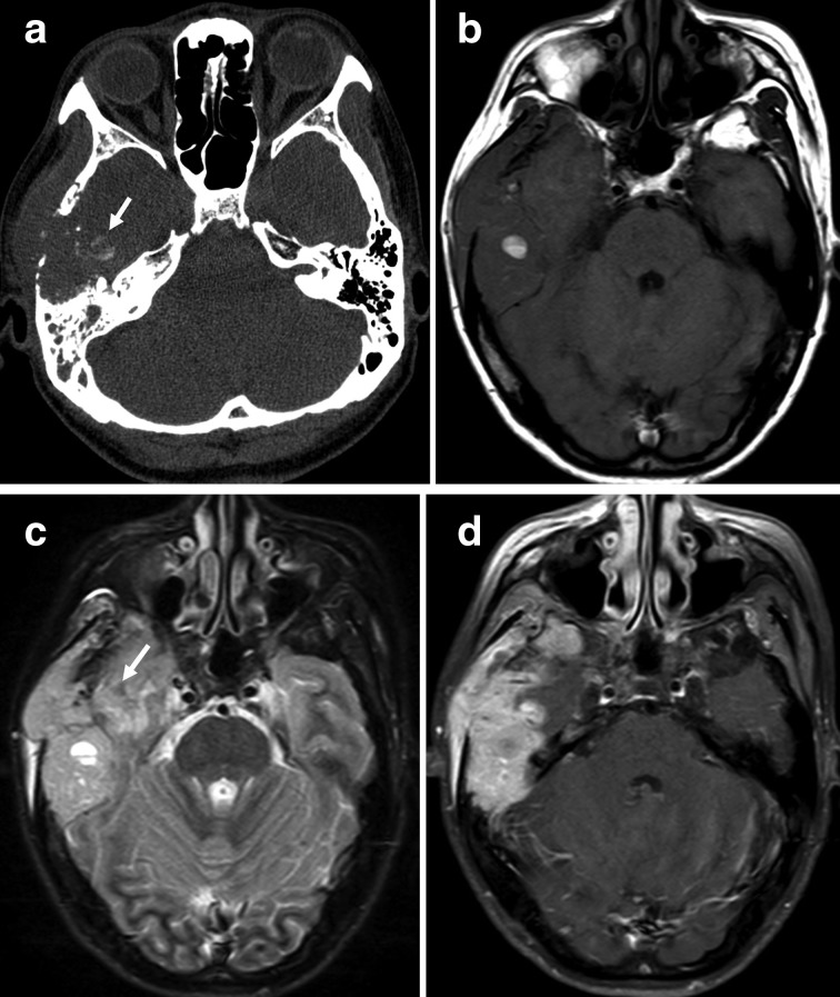
Figure 3. .
(a–d) A 40-year-old female with osteosarcoma of the right temporal bone (histological subtypes, osteoblastic). (a) Axial CT with the bone algorithm showing a lytic lesion of the right temporal bone with extraosseous soft tissue extension, suggestive of matrix mineralisation (white arrows). (b) Axial T1 weighted image of the soft tissue component showing heterogeneous signal intensity with patchy high signal intensity, suggestive of haemorrhage (white arrows). (c) FLAIR image of the soft tissue component showing heterogeneous high signal intensity with patchy low signal intensity, suggestive of matrix mineralization (white arrow). (d) Axial T1 weighted contrast-enhanced image showing a heterogeneous markedly enhancing mass in the right temporal bone with a lesion invading the temporal lobe.

