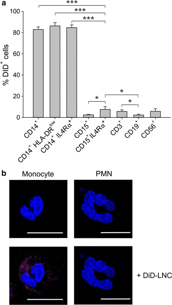
Fig. 3.

Uptake of neutral LNCs by leukocyte subsets in the peripheral blood of GBM patients. a Mean and SE of PBLs from 7 GBM patients incubated with 100 nm neutral LNCs for 3 h at a DiD concentration of 50 ng/ml. DiD-LNC uptake was evaluated by flow cytometry. Mann–Whitney test was performed, *P ≤ 0.05; **P ≤ 0.01; ***P ≤ 0.001. b PBMCs and PMNs isolated from GBM patients were incubated with 100 nm neutral LNCs for 3 h at a DiD concentration of 50 ng/ml. Then cells were washed and plated, nuclei were counterstained with DAPI, and the slides were analyzed by confocal microscopy. Representative fluorescence images (monocyte and PMN) are shown at a magnification of 150X. Cell size is reported by scale bar (10 µm)
