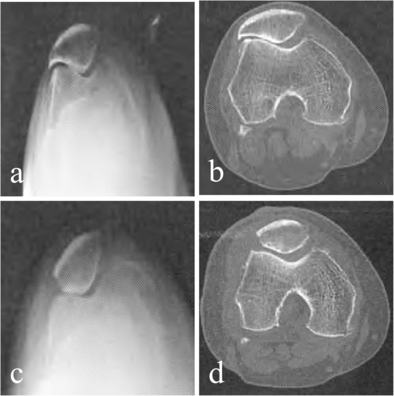
Fig. 2.

Comparison of preoperative and postoperative radiographic data of X-ray and CT examination of the ELPS patient. a, b Preoperative tangential X-ray imaging and CT tomography showed hook-shaped patella; the lateral space of the patellofemoral joint was significantly narrow and osteophyte formation was obvious. c, d Postoperative tangential X-ray imaging and CT tomography showed significant widening of the lateral space of the patellofemoral joint and its medial and lateral space restored to balance; the lateral hook-shaped patella had been shaped into a V-shaped patella; the osteophyte around the lateral patellofemoral joint had been removed and the joint returned to normal
