Fig. 5.
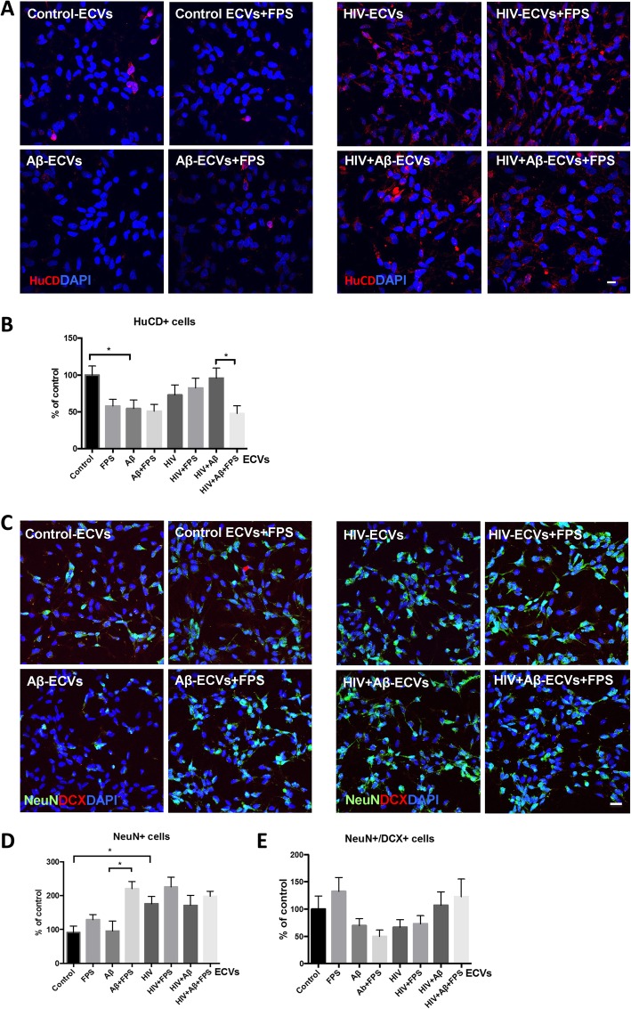
Impact of ECVs on NPC differentiation. HBMEC were treated with HIV and/or Aβ, and ECVs were isolated as in Fig. 1. NPC were differentiated for 3 days in the presence of HBMEC-derived ECVs. At the beginning of differentiation, selected NPC cultures were pretreated with 500 nM FPS-ZM1 (FPS) for 2 h followed by cotreatment with the isolated ECVs. At the end of the 3-day differentiation, confocal microscopy was performed for neuronal markers. Neuronal differentiation was assessed by counting Hu C/D-, NeuN- and doublecortin (DCX) positive cells. At least 9 images for every experimental condition from different samples were randomly acquired. Scale bar: 20 μm. a Representative images of Hu C/D immunoreactivity (red); nuclei are stained with DAPI (blue). b Hu C/D positive cells were counted from confocal microscopy images. c Representative images of NeuN (green) and doublecortin (DCX, red) immunoreactivity; nuclei are stained with DAPI (blue). d NeuN positive and e NeuN/DCX double positive cells were counted from confocal microscopy images. Values are mean ± SEM, n = 30–43 (Hu C/D); n = 7–15 (NeuN); n = 13 (NeuN/DCX). *Statistically significant as compared to control at p < 0.05

