Fig. 1.
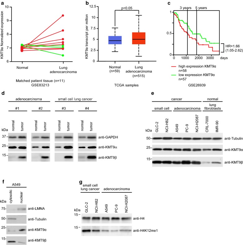
KMT9 is expressed in lung cancer tissue and cell lines. a Dynamics of KMT9α expression in matched normal and stage 1a lung adenocarcinoma tissue from eleven patients that underwent curative lobectomy. Normal samples were taken at 6 cm distance from macroscopic tumor sites. Data were extracted from (GSE83213). Red lines indicate increased expression of KMT9α in tumor (n = 8), green lines indicate decreased expression of KMT9α in tumor (n = 3). b TCGA data comparing KMT9α expression in n = 515 lung adenocarcinoma with non-matched normal lung tissue (n = 59). Data represent interquartile range including minimum, 25th percentile, median, 75th percentile and maximum values. Significance was accessed by t test. c Kaplan–Meier survival analysis of patients with adenocarcinoma expressing high (n = 58) and low (n = 57) KMT9α. Data were extracted from GSE26939. HR = hazard ratio. d Western blots of matched tissue from normal and tumor samples from patients with adenocarcinoma (#1 and #2) or SCLC (#3 and #4). Western blots were performed with the indicated antibodies. e Expression levels of KMT9α and KMT9β in human cell lines from SCLC (GLC-2 and NCI-H82), adenocarcinoma (A549, PC-9 and NCI-H2087) and human immortalized normal lung fibroblasts (CRL-7000 and IMR-90) were analyzed by western blot using the indicated antibodies. f In A549 cells, KMT9α and KMT9β are present in both nuclear and cytoplasmic compartments. Western blots were performed with the indicated antibodies. g Levels of H4K12me1 in SCLC (GLC-2 and NCI-H82) and adenocarcinoma (A549, PC-9 and NCI-H2087) cells were analyzed by western blotting using the indicated antibodies

