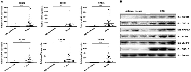
Figure 6.
Validation of six hub genes through experiments. (A) Validation of hub genes by reverse transcription-quantitative PCR. Boxplots indicate the medians and dispersions of 47 HCC and their adjacent tissue samples. P-values were determined using a Student' t-test. ***P<0.001 and ****P<0.0001 with comparisons shown by lines. (B) Western blotting detection of hub genes. Lysates from 3 pairs of HCC and adjacent tissues were subjected to western blotting with antibodies against CCNB2, CDC20, MAD2L1, MCM2, CENPF and BUB1B. β-actin was used as the reference gene. HCC, hepatocellular carcinoma; CCNB2, cyclin B2; CDC20, cell division cycle 20; MAD2L1, mitotic arrest deficient 2 like 1; MCM2 minichromosome maintenance complex component 2; CENPF, centromere protein F; BUB1B, BUB mitotic checkpoint serine/threonine kinase B.

