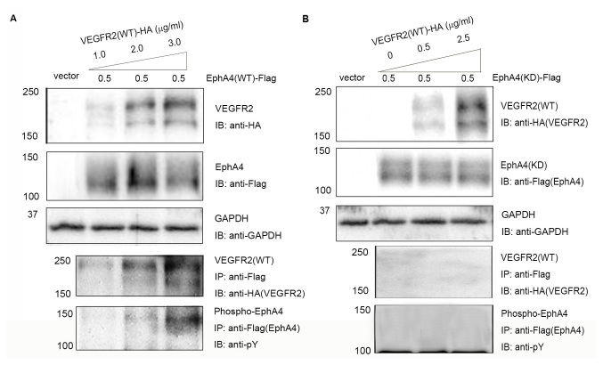
Figure 1.
Complex formation and transphosphorylation of EphA4 and VEGFR2 in transfected 293T cells. (A) 293T cells were co-transfected with pcDNA/EphA4-Flag (0.5 µg/ml) and increasing concentrations (1.0, 2.0 and 3.0 µg/ml) pcDNA/VEGFR2-HA. (B) 293T cells were co-transfected with pcDNA/EphA4(KD)-Flag (0.5 µg/ml) and increasing concentrations (0, 0.5 and 2.5 µg/ml) of pcDNA/VEGFR2(WT)-HA. Interactions were detected using SDS-PAGE and IB using anti-Flag antibodies following with IP using anti-HA antibodies. Tyrosine phosphorylation of EphA4 was detected using immunoprecipitation with anti-Flag antibodies followed by immunoblotting with anti-pY anitbodies. Eph, ephrin receptor; HA, hemagglutinin; IB, immunoblotting; IP, immunoprecipitation; KD, kinase-dead; pY, phosphotyrosine; VEGFR, vascular endothelial growth factor receptor; WT, wild-type.

