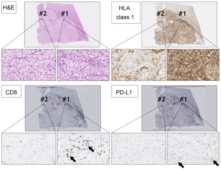
Figure 5.
Histological analysis of the primary RCC lesion. RCC sections were stained with H&E, anti-CD8 antibody (arrows), anti-HLA class 1 antibody, and anti-PD-L1 antibody (arrows). Magnification, ×2 or ×400. There were two staining patterns in RCC lesions #1 and #2. RCC, renal cell carcinoma; H&E, hematoxylin and eosin; HLA, human leukocyte antigen; PD-L1, programmed cell death ligand 1.

