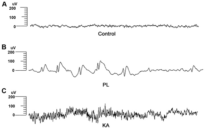
Figure 1.
Electroencephalograph recordings. (A) Rats in the control group showed normal brain activity. (B) A sharp and slow wave rhythm was observed in the PL group during myoclonic seizure attacks. (C) Multi-spike waves were recorded in the KA group during tonic-clonic seizure attacks. PL, pilocarpine; KA, kainic acid.

