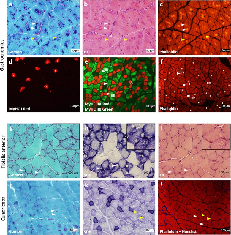
Fig. 2.
Histology and immunostaining of different skeletal muscles from NebY2303H,Y935X mice demonstrate multiple pathological features. a-c Nemaline bodies (white arrowheads; purple staining in Gomori, and intense staining with TRITC-phalloidin) in serial cross-sections of gastrocnemius (9-month-old male) stained with Gomori trichrome (a), H&E (b) and TRITC-phalloidin (c). Tubular aggregates (yellow arrowheads; pink in Gomori, negative with phalloidin) are a non-specific finding in older male mice from certain inbred strains. d-e Fibre typing was performed on serial sections using MyHC I (d), and MyHC IIA and IIB (e), antibodies. f TRITC-phalloidin visualised the actin-containing nemaline bodies most commonly locating in the fast MyHC type IIB fibres. All myofibres containing definite nemaline bodies are indicated with an asterisk (*), and 25/25 of these fibres are MyHC type IIB. Nemaline bodies were occasionally found in MyHC IIA fibres, however, no nemaline bodies were found in MyHC type I (slow) fibres (all type I fibres are indicated with an “I”). g-i Cross-sections of tibialis anterior (12-month-old female) stained with Gomori trichrome (g), SDH (h), and H&E (i), showing core-like structures in several myofibres (white arrowheads and inset). j-l Cross-sections of quadriceps (9 month male) stained with Gomori trichrome (j), SDH (k), and TRITC-phalloidin with Hoechst (l) showing internal nuclei (white arrowheads), split fibres (yellow arrowheads) and an occasional core-like structure (*)

