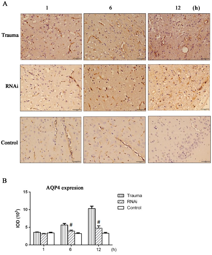
Figure 2.
Immunohistochemical staining of AQP-4. Brain tissue was harvested and sections were prepared for immunohistochemical staining. (A) AQP-4 immunohistochemical staining (magnification, ×400). (B) The results of immunohistochemistry staining were quantified in IOD. The data are presented as the mean ± standard deviation from at least three independent experiments. #P<0.05 vs. trauma group. AQP-4, aquaporin-4; IOD, integrated optical density; RNAi, RNA interference.

