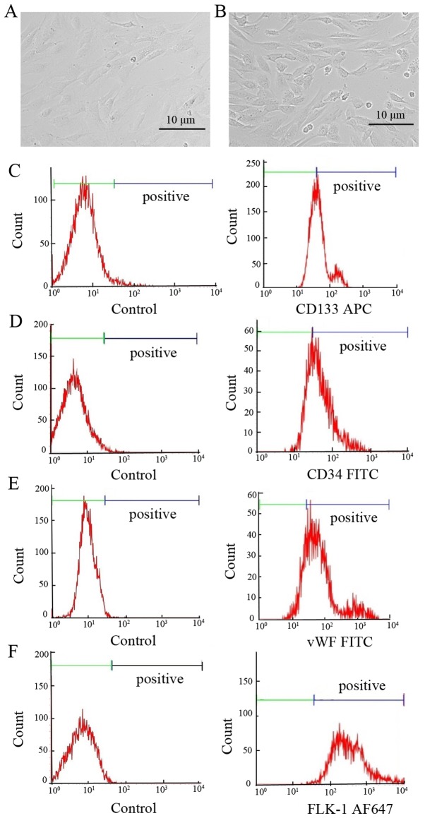
Figure 1.
Culture and identification of EPCs. (A and B) Morphology of EPCs after (A) 4 days and (B) 7 days of culture, assessed under a light microscope (scale bar, 10 µm); (C-F) EPCs were identified by flow cytometry following labeling with antibodies against (C) CD133, (D) CD34, (E) vWF and (F) Flk-1; isotype IgG served as a negative control (negative controls are presented in the left-hand panels and EPCs in the right-hand panels). EPC, endothelial progenitor cell; vWF, von Willebrand factor; Flk-1, fetal liver kinase 1.

