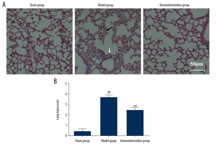
Figure 2.
Histological analysis of the rat lung tissues in the three study groups, the sham group, the model group, and the dexmedetomidine-treated group. (A) Lung injury was detected histologically. Hematoxylin and eosin (H&E). (Scale bar=50 μm). (B) Semiquantitative analysis of lung injury using histological scores. ** P<0.01 compared with the model group, ## P<0.01 compared with the sham group.

