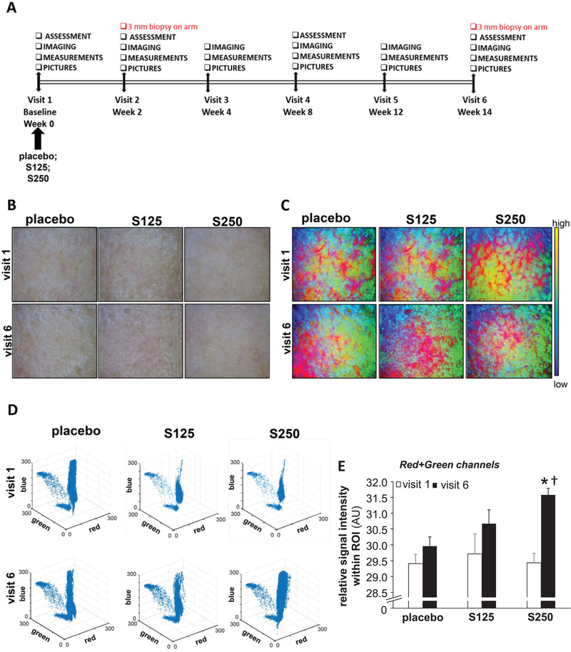
Figure 1.
Shilajit improves skin microperfusion. A, Study design. B, Dermascopic images of the cheek. C, MATLAB multicolor coded dermascopic images. D, 3-D scatterplot of the Visible Bands of MATLAB processed dermascopic images. E, The sum of the area under the curve of red and green channels were plotted graphically. The intensity of the red and green channels was calculated from the multicolor images processed by MATLAB software from the raw dermascopic images. S125 represents shilajit 125 mg and S250 represents shilajit 250 mg. Data are mean ± SEM (n = 13). *p < 0.05 compared to the baseline visit. †p < 0.05 compared to placebo.

