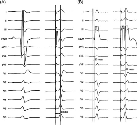
Figure 4.

The Stim‐LVAT of tip‐paced ECGs in patients with LBBP with and without LBB potential. One patient without LBB potential (A, left), had Stim‐LVAT of 68 ms (A, right). The other patient recorded with LBB potential (B, left), had Stim‐LVAT of 67 ms (B, right). ECG, echocardiography; LBB, left bundle branch; Stim‐LVAT, the interval from the pacing stimulus to the peak of the R‐wave
